KISS1R / neuroendocrine research · Research file
Kisspeptin-10
A source-linked five-part research file on KP-10 identity, KISS1R signaling, study records, comparisons, and evidence boundaries.
Five linked editorial lenses · research context only · sources linked below

The KISS1 gene encodes a precursor that can be processed into kisspeptin peptides. Early ligand-identification work connected kisspeptins with the receptor now called KISS1R, also known in the early literature as GPR54. KP-10 is commonly used as a short research ligand in receptor and signaling experiments.
Names, precursor numbering, and peptide forms should be checked against each original paper. A literature overview does not establish the identity, purity, stability, or other analytical characteristics of a particular research material.
- Research name: Kisspeptin-10 / KP-10
- Receptor terminology: KISS1R; early papers use GPR54
- Verify the exact peptide form and test article in each source
Source file
- Ohtaki et al. · Metastasis suppressor gene KiSS-1 encodes peptide ligand of a G-protein-coupled receptor · Nature (2001) ↗
- Kotani et al. · The metastasis suppressor gene KiSS-1 encodes kisspeptins, the natural ligands of the orphan G protein-coupled receptor GPR54 · Journal of Biological Chemistry (2001) ↗
- Oakley et al. · Kisspeptin signaling in the brain · Endocrine Reviews (2009) ↗

In commonly described models, receptor activation is associated with PLC signaling, generation of IP3 and DAG, and intracellular calcium mobilization. Additional signaling observations appear across different experimental systems. The details should be attributed to the specific assay and cell model rather than presented as one universal pathway diagram.
For any proposed mechanism, check whether the experiment directly tested the receptor, the second messenger, and the downstream response. An illustration of a pathway is not itself evidence of a binding pose, receptor occupancy, or a complete causal sequence.
- Distinguish KISS1R/GPR54 from downstream signaling components
- Track the cell system, receptor construct, assay, and measured signal
- Keep inferred pathway steps separate from directly tested observations
Source file
- Pinilla et al. · Kisspeptins and reproduction: physiological roles and regulatory mechanisms · Physiological Reviews (2012) ↗
- Oakley et al. · Kisspeptin signaling in the brain · Endocrine Reviews (2009) ↗
- Kotani et al. · The metastasis suppressor gene KiSS-1 encodes kisspeptins, the natural ligands of the orphan G protein-coupled receptor GPR54 · Journal of Biological Chemistry (2001) ↗

Early receptor studies established the kisspeptin–GPR54 ligand relationship. Later reviews summarize assays using cell systems and discuss intracellular signaling readouts. Those reviews are useful maps to the primary record, but their summaries should not replace the original methods, controls, and data.
Animal studies compare peptide forms and measure defined endpoints under specific experimental conditions. Read the test article, species, route, timing, comparator, and measured endpoint from the original report before comparing its findings with cell-based receptor assays.
- Ohtaki and Kotani (2001): foundational ligand/receptor identification work
- Reviews summarize receptor and second-messenger experiments across models
- Animal-model observations remain specific to their design and measured endpoints
Source file
- Ohtaki et al. · Metastasis suppressor gene KiSS-1 encodes peptide ligand of a G-protein-coupled receptor · Nature (2001) ↗
- Kotani et al. · The metastasis suppressor gene KiSS-1 encodes kisspeptins, the natural ligands of the orphan G protein-coupled receptor GPR54 · Journal of Biological Chemistry (2001) ↗
- Pinilla et al. · Kisspeptins and reproduction: physiological roles and regulatory mechanisms · Physiological Reviews (2012) ↗
- Thompson et al. · Differential effects of kisspeptin-10 and kisspeptin-54 on gonadotropin secretion in rats · American Journal of Physiology-Endocrinology and Metabolism (2006) ↗
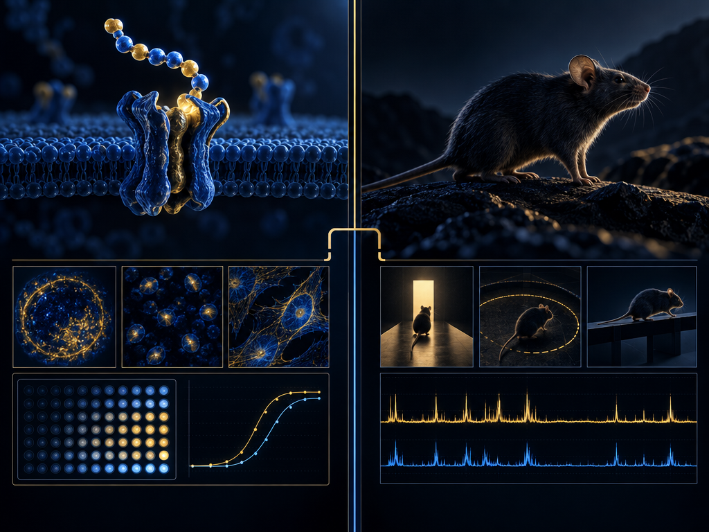
A published rat study compared KP-10 and the longer kisspeptin-54 in a specific experimental setting and reported differences in measured responses. Such findings cannot be separated from the study's design, peptide handling, model, timing, and endpoint, nor treated as a universal ranking of the peptide forms.
Newer work also explores kisspeptin signaling in other cell types, including a 2024 study of osteoclast biology. That is a distinct research context from neuroendocrine receptor studies. Separate results should not be merged into one mechanism or treated as evidence that different models are directly comparable.
- Compare KP-10 with longer forms only within the cited experimental design
- Distinguish neuroendocrine assays from other cell-type research
- A cross-study contrast is not a direct head-to-head result unless the design is
Source file
- Thompson et al. · Differential effects of kisspeptin-10 and kisspeptin-54 on gonadotropin secretion in rats · American Journal of Physiology-Endocrinology and Metabolism (2006) ↗
- Uenoyama et al. · Kisspeptin signaling in osteoclasts and bone biology · Nature Communications (2024) ↗
- Pinilla et al. · Kisspeptins and reproduction: physiological roles and regulatory mechanisms · Physiological Reviews (2012) ↗

The source record spans ligand identification, receptor pharmacology, reviews of signaling, animal experiments, and newer work in other cell types. These sources do not all test the same question. Receptor activity in a defined assay does not by itself establish every downstream step or show that an observation transfers across tissues, species, or experimental conditions.
To evaluate a claim, follow the citation to the original design, determine what was directly measured, and record the limitations and competing interpretations. A review can orient the reader; primary methods and results determine the scope of a specific finding.
- Receptor pharmacology is not interchangeable with a whole-model outcome
- Peptide form, stability, species, and experimental conditions affect comparisons
- Do not generalize a result beyond its tested model and endpoint
- Unresolved mechanisms should remain open questions
Source file
- Ohtaki et al. · Metastasis suppressor gene KiSS-1 encodes peptide ligand of a G-protein-coupled receptor · Nature (2001) ↗
- Kotani et al. · The metastasis suppressor gene KiSS-1 encodes kisspeptins, the natural ligands of the orphan G protein-coupled receptor GPR54 · Journal of Biological Chemistry (2001) ↗
- Pinilla et al. · Kisspeptins and reproduction: physiological roles and regulatory mechanisms · Physiological Reviews (2012) ↗
- Oakley et al. · Kisspeptin signaling in the brain · Endocrine Reviews (2009) ↗
- Thompson et al. · Differential effects of kisspeptin-10 and kisspeptin-54 on gonadotropin secretion in rats · American Journal of Physiology-Endocrinology and Metabolism (2006) ↗
- Uenoyama et al. · Kisspeptin signaling in osteoclasts and bone biology · Nature Communications (2024) ↗
Reading the source record
Follow each link to the original methods, endpoints, and limitations. This editorial summary is not a substitute for the source paper; interpretations should remain within the tested design and evidence scope.